
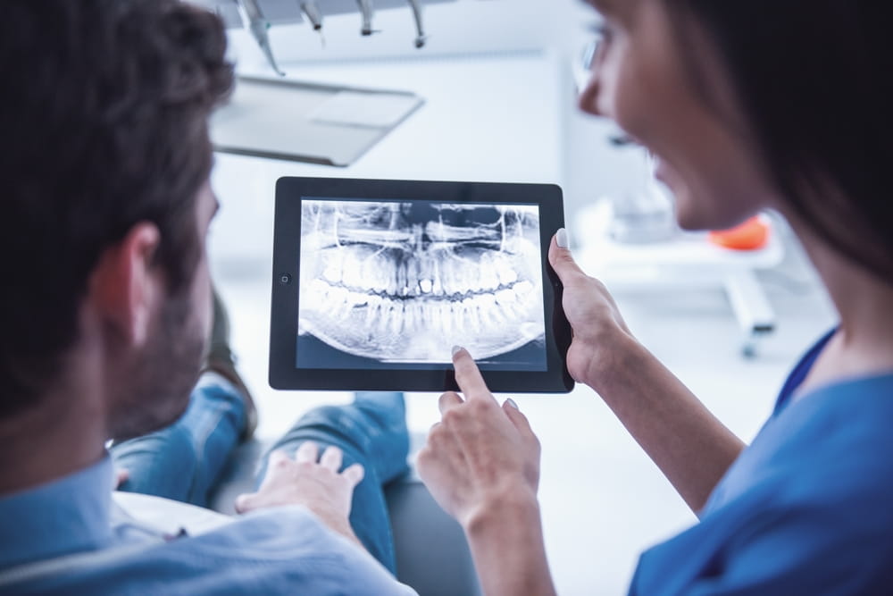
At Wyngate Dental of Bethesda, advances in dental technology continue to improve the way oral health conditions are detected, diagnosed, and monitored. Among the most significant innovations in modern dentistry is the use of digital X-rays. While traditional film-based radiographs have been used successfully for decades, digital imaging has transformed diagnostic capabilities by providing clearer images, faster results, and greater precision.
Diagnostic accuracy plays a critical role in successful dental care. The earlier a problem is identified, the more treatment options may be available and the better the chances of preserving oral health. Digital X-rays have become an important tool in helping dental professionals identify issues that may not be visible during a visual examination alone, allowing patients to receive more timely and effective care.
Why Dental X-Rays Are Essential for Oral Health
Even the most thorough clinical examination cannot reveal everything happening beneath the surface of the teeth and gums. Many dental conditions develop in areas that cannot be seen directly, including between teeth, beneath existing restorations, within the jawbone, or below the gumline. Dental X-rays provide valuable insight into these hidden structures.
Without radiographic imaging, conditions such as cavities, bone loss, impacted teeth, infections, and developing abnormalities may go undetected until they become more advanced. By providing a detailed view of the underlying anatomy, X-rays help clinicians identify concerns before they cause significant symptoms or damage.
Regular imaging is often an important component of preventive dental care. Combined with routine examinations and services such as Cleaning and Hygiene, digital X-rays support a more complete understanding of a patient’s oral health status.
What Makes Digital X-Rays Different?
Digital X-rays utilize electronic sensors rather than traditional photographic film. When an image is captured, it appears almost instantly on a computer screen, eliminating the need for chemical processing and lengthy development times. This immediate access allows dental professionals to review images during the appointment and discuss findings with patients in real time.
One of the most important advantages of digital technology is image quality. Digital radiographs often provide sharper, more detailed images that can be enlarged and adjusted for contrast, brightness, and clarity. These enhancements allow clinicians to examine areas of concern more closely and identify subtle abnormalities that may be difficult to detect on conventional film.
Digital systems also create a more efficient workflow. Images can be securely stored, compared over time, and shared with specialists when needed, helping support coordinated and comprehensive patient care.
Enhanced Detection of Dental Conditions
The improved image quality offered by digital X-rays contributes directly to greater diagnostic accuracy. High-resolution imaging allows clinicians to identify small changes in tooth structure and surrounding tissues that may indicate developing problems.
Digital X-rays can help detect:
- Early tooth decay between teeth
- Bone loss associated with periodontal disease
- Dental abscesses and infections
- Cracks and fractures in teeth
- Impacted or unerupted teeth
- Changes in jawbone structure
- Failing restorations beneath crowns or fillings
- Developmental abnormalities
The ability to identify these issues at earlier stages often leads to less invasive treatment options. For example, a small cavity discovered through radiographic imaging may be treated with a simple Tooth Colored Filling, whereas an undetected cavity could eventually require more extensive restorative care.
Supporting More Accurate Treatment Planning
Digital X-rays do more than diagnose existing conditions—they also help guide treatment decisions. Accurate imaging allows dental professionals to assess the severity and extent of a problem before recommending a treatment plan. This information supports more predictable outcomes and helps patients better understand their oral health needs.
For example, when evaluating candidates for Dental Implants, clinicians rely on imaging to assess bone levels and determine whether sufficient support exists for implant placement. Similarly, digital radiographs play an important role in periodontal evaluations, helping identify bone loss patterns and supporting treatments such as Arestin or scaling and root planing.
The ability to monitor changes over time is another significant advantage. By comparing current images with previous radiographs, clinicians can track disease progression, evaluate treatment effectiveness, and identify emerging concerns before they become serious.
Improved Safety and Patient Experience
One of the most widely recognized benefits of digital X-rays is reduced radiation exposure. Modern digital systems generally require significantly less radiation than traditional film-based radiography while still producing highly detailed images. This reduction contributes to a safer diagnostic process for patients of all ages.
The patient experience is also enhanced through greater convenience and comfort. Digital sensors provide immediate results, reducing appointment times and eliminating the waiting period associated with film processing. Patients can view their images alongside their provider, making it easier to understand diagnoses and treatment recommendations.
Additionally, digital imaging eliminates the need for chemical processing solutions used in traditional film development, making it a more environmentally friendly option for dental practices.
The Future of Precision Dentistry
As dental technology continues to advance, digital imaging remains a cornerstone of modern diagnostic dentistry. High-quality images, enhanced visualization tools, reduced radiation exposure, and streamlined workflows all contribute to improved diagnostic precision and patient care. The ability to identify oral health concerns earlier allows for more proactive treatment and better long-term outcomes.
At Wyngate Dental of Bethesda, patient education is an important part of promoting lifelong oral health. Understanding how digital X-rays improve diagnostic accuracy helps patients appreciate the role technology plays in detecting problems early, guiding treatment decisions, and supporting healthier smiles. With the aid of advanced imaging, dental professionals can provide more informed evaluations and help patients maintain optimal oral health for years to come.
Resources
White, S. C., & Pharoah, M. J. (2014). Oral Radiology: Principles and Interpretation. Elsevier Health Sciences.
Farman, A. G., & Farman, T. T. (2005). A Comparison of 18 Different X-Ray Detectors Currently Used in Dentistry. Oral Surgery, Oral Medicine, Oral Pathology, Oral Radiology, and Endodontology.
Iannucci, J. M., & Howerton, L. J. (2021). Dental Radiography: Principles and Techniques. Elsevier.